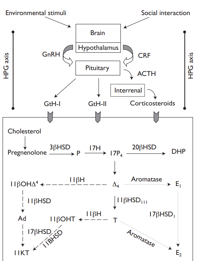
Vertebrates of both sexes possess a germinal epithelium consisting of germ and somatic cells. The gonads are mesodermal in origin and develop in close association with the nephric system. In elasmobranchs a lateral cortex gives rise to the ovary, and a more medial area becomes the testis. One of these usually atrophies early in development when the sex is determined. In cyclostomes and teleosts the gonads derive from a single region equivalent to the cortex. While most fishes have paired gonads, fusion of two primordia in the cortex of lampreys leads to a single gonad, whereas in the hagfishes one gonad fails to develop, and there is only a single ovary in some sharks (Lagler et al., 1962).
The gametes first appear as primordial germ cells, the maturation of which is regulated by hormones. The ovary is usually hollow, although the wall may become folded as its internal surface area increases.The oogonia become surrounded by a single layer of follicular cells in cyclostomes and teleosts but a multilayer in elasmobranchs. In the primary oocyte stage the developing eggs are supplied with yolk by the follicular cells during vitellogenesis. The yolk attains a granular texture in most fishes; however in atherinomorph fishes it retains a liquid composition. As will be noted below, this is not the only reproductive peculiarity of the Atherinomorpha. At the completion of maturation the oocyte becomes free of the follicle during ovulation and water is taken up, causing the egg to swell. In batch spawners, populations of different-sized oocytes can be seen, each corresponding to a future batch of eggs for release. In some species the mature eggs are released into the body cavity and pass into the funnel-shaped opening of the oviduct and so to the exterior; in other species the ovarian lumen and oviduct are continuous.

Elasmobranch and bony fish testes are unique among vertebrates in that spermatocyte development occurs in cysts (spermatocysts) that contain spermatocytes of a single genetic lineage (Miura and Miura, 2003). During development, spermatocytes migrate from their site of origin and empty eventually into an efferent duct. Despite this similarity, the teleost and elasmobranch testes are differently organized. In most sharks the testes are paired cylindrical organs with a ridge of germinal tissue running along each lateral surface. Spermatocyst development proceeds across (diametrically) each testis leading toward the medially located efferent ducts. In contrast to this pattern, the testes of lamniform sharks are cylindrical organs with several germinal zones surrounded by seminiferous follicles. Spermatocyte development occurs radially, with the mature spermatozoa being released in efferent ducts that surround each group of follicles (Jones and Jones, 1982). Skates and rays appear to have a different structure termed “compound” that shows characteristics of both other types (Pratt, 1988).
As described by Parenti and Grier (2004), bony fishes show even greater variation of testis morphology. Latimeria, non-teleost Actinopteryigii, and basal teleosts are characterized by testes with anastomosing tubules (although it must be noted that these are fundamentally different from the tubular testes of amniotic vertebrates) whereas derived taxa (Neoteleosts) have a lobular testes, which in turn can be divided into a so-called “perciform” testes (because it was first observed among perciform fishes, but in fact is also found in all major groups except Atherinomorphs) in which spermatocytes are distributed along the entire length of the lobules and an “atherinomorph” testis (which is restricted to and found universally among atherinomorph taxa) in which spermatocytes are restricted to the distal ends of the lobules.
The spermatogonia pass through a spermatocyte and spermatid stage before becoming spermatozoa. During spawning these pass directly to the exterior via the seminiferous tubules and vas deferens. En route the spermatozoa are diluted with secretions of seminal fluid. Some elasmobranchs, for instance the basking shark, and live-bearing teleosts produce spermatophores or packets of spermatozoa.


