The released egg is protected by a fairly tough chorion or egg case. Within this the cytoplasm and yolk are contained by a vitelline membrane. Often one or more oil globules are present. Fertilization occurs by a spermatozoon passing through a funnel-shaped micropyle leading to a fusion of the pronuclei of the sperm and egg. This leads to activation when the oocyte, arrested before fertilization, then resumes development. The vitelline membrane separates from the chorion creating a perivitelline space, and the micropyle is plugged, preventing further spermatozoa entering.
Some elas mobranchs have polyspermy in which a number of spermatozoa may enter the micropyle but only one fuses with the nucleus of the egg, the rest being resorbed and perhaps used as an additional nutrient. In salmonids, water activation takes place as the egg absorbs water regardless of whether fertilization takes place or not. In river water, the vitelline membrane becomes opaque and its permeability changes. If spermatozoa are not present, the fertilizability of the egg is soon lost. After fertilization the chorion hardens, protecting the egg. This is especially valuable in waves or surf or if the eggs are buried in gravel. The chorion remains permeable to water and small molecules but the site of osmoregulation is the vitelline membrane.
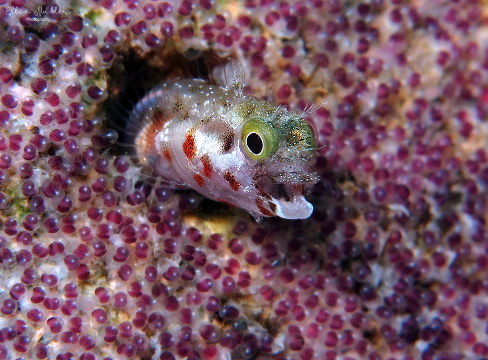
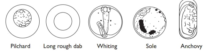
Fish eggs are usually round although the hagfish (Myxine), anchovy Engraulis) and bitterling (Rhodeus) have ovoid eggs, and in some gobies they are pear-shaped. Most species are telolecithal with yolk concentrated at the vegetative pole and the cytoplasm at the animal pole giving a “polarity” to the egg. The egg goes through a process of cleavage and morphogenesis as the cells divide, which form layers, and then organs. In lampreys cleavage is holoblastic, the entire egg dividing to form smaller or micromeres at the animal pole and macromeres at the vegetative pole. In hagfish, elasmobranchs and teleosts development is meroblastic. Here, cleavage at the animal pole leads to a blastoderm or cap of cells. The blastoderm overgrows the yolk (epiboly) eventually enclosing it to form a gastrula, a hollow sphere of cells containing yolk with a small opening into the perivitelline space – the blastopore.
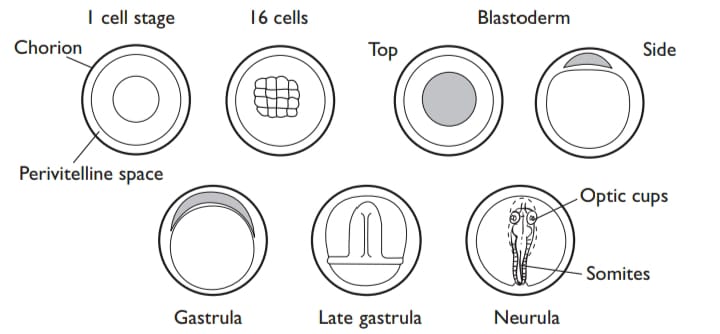
The embryonic axis is laid down in relation to the dorsal lip of the blastopore and the neurula stage forms with the future head, spinal cord, and body musculature soon visible. After a time, the tail region grows away from the neurula and coils around inside the perivitelline space. The optic cups (the future eyes) and heart are the first organs to be identified easily. The heart starts to beat well before hatching and in some species the eyes become pigmented and functional, and the embyonic fish often wriggles and rotates within the chorion. The prelude to hatching is a softening of the chorion as a result of enzymes secreted by glands on the head. Some of the chorionic material is probably utilized as a nutrient by the embryo before hatching. The embryo then breaks free from the chorion to become a larva.