Trematodes are all parasitic flukes, and as adults they are almost all found as endoparasites of vertebrates. They are chiefly leaf like in form with one or more suckers but lack the opisthaptor present in monogenean fluke Other structural adaptations for parasitism are apparent: various penetration glands or glands to produce cyst material, organs for adhesion such as suckers and hooks, and increased reproductive capacity. Otherwise, trematodes share several characteristics with ectolecithal turbellarians, such as a well-developed alimentary canal (but with the mouth at the anterior, or cephalic, end) and similar reproductive, excretory, and nervous systems, as well as a musculature and parenchyma that are only slightly modified from those of turbellarians. Sense organs are poorly developed.
Of the subclasses of Trematoda, subclass Aspidogastrea is the least well known. Most parasites in this group have only a single host, usually a mollusc. If there is a second host, it is usually a fish or turtle. Subclass Digenea (Gr. dis, double, + genos, race) is the largest and best known, with many species of medical and economic importance.
With rare exceptions, digeneans have a complex life cycle, the first (intermediate) host being a mollusc and the definitive host (the host in which sexual reproduction occurs, sometimes called the final host) being a vertebrate. In some species a second, and sometimes even a third, intermediate host intervenes. The group has radiated greatly, and its members parasitize almost all kinds of vertebrate hosts. Digeneans inhabit, according to species, a wide variety of sites in their hosts: all parts of the digestive tract, respiratory tract, circulatory system, urinary tract, and reproductive tract.
Among the world’s most amazing biological phenomena are digenean life cycles. Although cycles of different species vary widely in detail, a typical example would include an adult, egg (shelled embryo), miracidium, sporocyst, redia, cercaria, and metacercaria stages . The shelled embryo or larva usually passes from the definitive host in excreta and must reach water to develop further. There, it hatches to a free-swimming, ciliated larva, the miracidium. The miracidium penetrates the tissues of a snail, where it transforms into a sporocyst. Sporocysts reproduce asexually to yield either more sporocysts or a number of rediae. Rediae, in turn, reproduce asexually to produce more rediae or to produce cercariae. In this way a single egg can give rise to an enormous number of progeny. Cercariae emerge from the snail and can either penetrate the final host directly (for example, the blood fluke Schistosoma mansoni), penetrate a second intermediate host (for example, the lung fluke Paragonimus westermani), or encyst on aquatic vegetation (for example, the intestinal fluke Fasciolopsis buski). At this point, cercariae develop into metacercariae, which are essentially juvenile flukes. When the metacercariae are eaten by the final host, the juveniles migrate to the site of final infection and grow into adults.
Some of the most serious parasites of humans and domestic animals belong to Digenea . The first digenean life cycle revealed was that of Fasciola hepatica (L. fasciola, a small bundle, band), which causes “liver rot” in sheep and other ruminants. Adult flukes live in the bile passage of the liver, and eggs are passed in feces. After hatching, a miracidium penetrates a snail to become a sporocyst. There are two generations of rediae, and the cercaria encysts on vegetation. When infested vegetation is eaten by a sheep or other ruminant (or sometimes humans), the metacercariae excyst and grow into young flukes.

Clo-norchis (Gr. clon, branch, orchis, testis) is the most important liver fluke of humans and is common in many regions of eastern Asia, especially in China, Southeast Asia, and Japan. Cats, dogs, and pigs are also often infected.The worms vary from 10 to 20 mm in length. Their structure is typical of many trematodes in most respects. They have an oral sucker and a ventral sucker. The Digestive system consists of a pharynx, a muscular esophagus, and two long, unbranched intestinal ceca. The excretory system consists of two protonephridial tubules, with branches lined with flame cells. The two tubules unite to form a single median bladder that opens to the outside. The nervous system, like that of other flatworms, consists of two cerebral ganglia connected to longitudinal cords that have transverse connectives.

As is common in flukes, about 80% of the body is devoted to reproduction. The reproductive system is hermaphroditic and complex. They have two branched testes that unite to form a single vas deferens, which widens into a seminal vesicle. The seminal vesicle leads into an ejaculatory duct, which terminates at the genital opening. The female system contains a branched ovary with a short oviduct, which is joined by ducts from the seminal receptacle and vitellaria at an ootype. The ootype is surrounded by a glandular mass, Mehlis’s gland, of uncertain function. From Mehlis’s gland the much-convoluted uterus runs to the genital pore. Cross-fertilization between individuals is usual, and sperm are stored in the seminal receptacle. When an oocyte is released from the ovary, it is joined by a sperm and a group of vitelline cells and is fertilized. The vitelline cells release a proteinaceous shell material, which is stabilized by a chemical reaction; the Mehlis’s gland secretions are added, and the egg passes into the uterus.
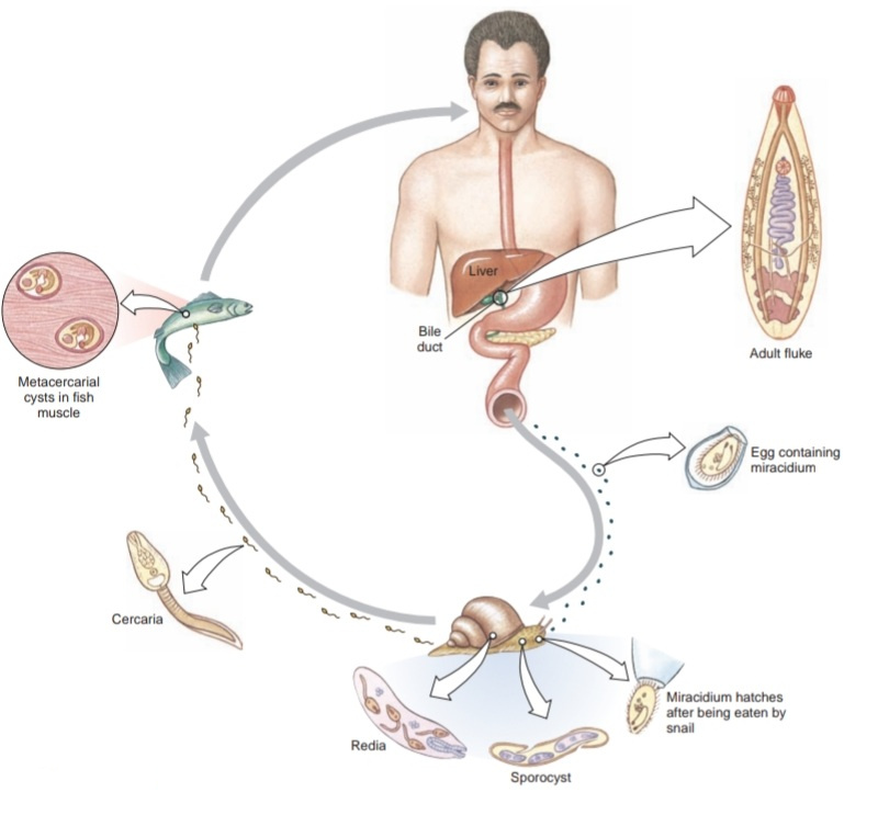
The normal habitat of the adults is in the bile passageways of humans and other fish-eating mammals . Eggs, each containing a complete miracidium, are shed into water with the feces but do not hatch until they are ingested by the snail Parafossarulus or related genera. Eggs, however, may live for some weeks in water. In a snail a miracidium enters the tissues and transforms into a sporocyst, which produces one generation of rediae. A redia is elongated, with an alimentary canal, a nervous system, an excretory system, and many germ cells in the process of development. Rediae pass into the liver of the snail where the germ cells continue embryonation and give rise to tadpolelike cercariae. These two asexual stages in the intermediate host allow a single miracidium to produce as many as 250,000 infective cercariae.
Cercariae escape into the water and swim until they encounter a fish of family Cyprinidae. They then bore under scales into the fish’s muscles. Here cercariae lose their tails and encyst as metacercariae. If a mammal eats raw or undercooked infected fish, the metacercarial cyst dissolves in the intestine, and young flukes apparently migrate up the bile duct, where they become adults. There the flukes may live for 15 to 30 years.
The effect of these flukes on a person depends mainly on the extent of the infection but includes abdominal pain and other abdominal symptoms. A heavy infection can cause a pronounced cirrhosis of the liver and death. Cases are diagnosed through fecal examinations. Destruction of snails that carry larval stages has been used as a method of control. However, the simplest method to avoid infection is to make sure that all fish used
as food is thoroughly cooked.
Useful External Links