One of the most interesting—and sometimes puzzling—aspects of this phylum is the dimorphism and often polymorphism displayed by many of its members. All cnidarian forms fit into one of two morphological types (dimorphism): a polyp, or hydroid form, which is adapted to a sedentary or sessile life, and a medusa, or jellyfish form, which is adapted for a floating or free-swimming existence. Superficially the polyp and medusa seem very different, but actually each has retained the saclike body plan characteristic of the phylum.
A medusa is essentially an unattached polyp with the tubular portion widened and flattened into a bell shape. Polyps Most polyps have tubular bodies. A mouth surrounded by tentacles defines the oral end of the body. The mouth leads into a blind gut or gastrovascular cavity. The aboral end of the polyp is usually attached to a substratum by a pedal disc or other device.

Polyps may reproduce asexually by budding, fission, or pedal laceration. In budding, a knob of tissue forms on the side of an existing polyp and develops a functional mouth and tentacles. If a bud detaches from the polyp that made it, a clone is formed. If a bud stays attached to the polyp that made it, a colony will form and food may be shared through a common gastrovascular cavity. Polyps that do not bud are solitary; others form clones or colonies. Thedistinction between colonies and clones is sometimes blurred when a colony fragments.
A shared gastrovascular cavity permits polyp specialization. Many colonies include several morphologically distinct polyps, each specialized for a certain function, such as feeding, reproduction, or defense. Such colonies exhibit polymorphism. In class Hydrozoa, feeding polyps or hydranths, are easily distinguished from reproductive polyps, or gonangia, by the absence of tentacles in gonangia. Gonangia typically make medusae. Other methods of asexual reproduction in polyps are fission, where an individual divides in half as one side of the polyp pulls away from the other side, or pedal laceration, where tissue torn from the pedal disc develops into tiny new polyps. Pedal laceration and fission are common in sea anemones in class Anthozoa.

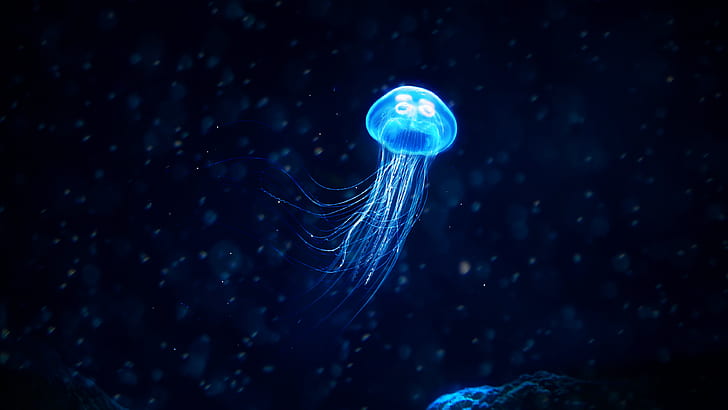
Medusae Medusae are usually free swimming and have bell or umbrella-shaped bodies. They often exhibit tetramerous symmetry where body parts are arranged in fours. The mouth is usually centered on the concave (subumbrellar) side, and it may be pulled downward into frilly lobes that extend a long way beneath the umbrella or bell. Tentacles extend outward from the rim of the umbrella. Medusae have sensory structures for orientation (statocysts) and light reception (ocelli). Sensory information is integrated with motor response by a nerve ring at the base of the bell; two such rings are present in hydrozoan medusae.
Medusae of class Scyphozoa are often called scyphomedusae, whereas those of class Hydrozoa are hydromedusae. Hydromedusae differ from scyphomedusae by the presence of a velum, a shelflike fold of tissue from the bottom of the bell that extends into the bell. By reducing the cross-sectional area at the bottom of the bell, the velum increases the exit velocity of water from the bell, making each pulsation more efficient.
Life Cycles
In a cnidarian life cycle, polyps and medusae play different roles. The particular sequence of forms in the life cycle varies among cnidarian classes, but in general, a zygote develops into a motile planula larva. The planula settles on a hard surface and metamorphoses into a polyp. A polyp may make other polyps asexually, but eventually it produces free-swimming medusae by asexual reproduction. Polyps produce medusae by budding, or other specialized methods like strobilation. Medusae reproduce sexually and are dioecious.
A life cycle that contains both an attached polyp and a swimming medusa permits organisms to take advantage of both pelagic (open water) and benthic (bottom) environments. Such life cycles occur in true jellyfishes of class Scyphozoa where the medusa is large and conspicuous and the polyps are typically very small. Most hydroids of class Hydrozoa also feature a sessile polyp stage, often in colonies, and a pelagic medusa stage. However, there are many variations on the typical pattern.
In some hydrozoans, the polyp colony is not sessile, but drifts across the ocean surface. The Portuguese man-of-war, Physalia, is one such drifter, using an inflated polyp as a gas-filled float. Other colonies are collections of both polyps and medusae where pulsating bells propel the colony through the water. Several life cycles do not include medusae. Anthozoans are presumed to have diverged from an ancestor of the other cnidarians before the medusa evolved in the latter branch, but other cnidarians, including the hydrozoan Hydra, probably lost the medusa secondarily. The mechanism of loss is not clear in Hydra, but in other hydrozoans, a pattern of loss can be inferred from a comparison of modern forms.
Most hydrozoans release medusae that later make gametes, but a few forms make medusae without releasing them from the colony. Gametes then form in the gonads of the medusae retained by the polyp colony. In some species only a short cuplike form surrounds the gonads, and in others gonads develop right on the polyp colony with no trace of a medusa body. The latter organisms likely represent an extreme form of medusa retention and reduction.
Body Wall
The cnidarian body comprises an outer epidermis, derived from ectoderm, and an inner gastrodermis, derived from endoderm, with mesoglea between them. The gastrodermis lines the gut cavity and functions mainly in digestion. In polyps of the solitary hydrozoan, Hydra, the epidermal layer contains several cell types, including epitheliomuscular, interstitial, gland, sensory, and nerve cells, as well as cnidocytes. Cnidarian bodies extend, contract, bend, and pulse, all in the absence of true mesodermally derived muscle cells.

Instead, epitheliomuscular cells form most of the epidermis and serve both for covering and for muscular contraction. The bases of most such cells are extended parallel to the tentacle or body axis and contain myofibrils; they form the functional equivalent of a layer of longitudinal muscle next to the mesoglea. Contraction of these fibrils shortens the body or tentacles. The mesoglea lies between the epidermis and the gastrodermis and is attached to both layers. It is gelatinous, or jellylike, and both epidermal and gastrodermal cells send processes into it. In polyps, it is a continuous layer extending over both body and tentacles, thinnest in the tentacles and thickest in the stalk portion. This arrangement allows the pedal region atthe base of the animal to withstand great mechanical strain and gives the tentacles more flexibility.
The mesoglea helps to support the body and acts as a type of elastic skeleton. In class Anthozoa, the mesoglea is substantial and possesses ameboid cells. The mesogleal layer is also very thick in scyphozoan medusae and contains ameboid cells and fibers. The medusa bell has a fairly firm consistency, despite the fact that mesogleal jelly is 95% to 96% water. The buoyant mass of mesogleal “jelly” gives medusae the common name jellyfishes. The mesoglea is much thinner in the bells of hydromedusae and lacks ameboid cells or fibers.
Cnidocytes
As the opening essay attests, many cnidarians are very effective predators on prey larger and more intelligent than themselves. Such efficient predation is made possible by amply arming the tentacles with a unique cell type, the cnidocyte. Cnidocytes are borne in invaginations of ectodermal cells and, in some forms, in endodermal cells. Each cnidocyte produces one of over 20 kinds of distinctive organelles called cnidae that are discharged from the cell. During its development, a cnidoctye is properly called a cnidoblast. Once its cnida has been discharged, a cnidocyte is absorbed and replaced.
One type of cnida, the nematocyst, is used to inject a toxin for prey capture and defense. Nematocysts are tiny capsules composed of material similar to chitin and containing a coiled tubular “thread” or filament, which is a continuation of the narrowed end of the capsule. This end of the capsule is covered by a little lid, or operculum. The inside of the undischarged thread may bear tiny barbs, or spines. Not all cnidae have barbs or inject poison. Some kinds, for example, do not penetrate prey but rapidly recoil like a spring after discharge, grasping and holding any part of the prey caught in the coil. Adhesive cnidae usually do not discharge in food capture, but are used in attachment and locomotion.

Except in Anthozoa, cnidocytes are equipped with a trigger like cnidocil, which is a modified cilium. Anthozoan cnidocytes have a somewhat different ciliary mechanoreceptor. In some sea anemones, and perhaps other cnidarians, small organic molecules from the prey “tune” the mechanoreceptors, sensitizing them to the frequency of vibration caused by prey swimming. Tactile stimulation causes the nematocyst to discharge.
The mechanism of nematocyst discharge is remarkable. Evidence indicates that discharge is due to a combination of tensional forces generated during nematocyst formation and to an astonishingly high osmotic pressure within the nematocyst: 140 atmospheres. When stimulated to discharge, the high internal osmotic pressure causes water to rush into the capsule. The operculum opens, and the rapidly increasing hydrostatic pressure within the
capsule forces the thread out with great force, turning inside out as it goes. At the everting end of the thread, the barbs flick to the outside like tiny switchblades. This minute but awesome weapon then injects poison when it penetrates prey.
Scyphomedusae are typically larger than hydromedusae, but their basic form is similar. The mouth edge is extended as a manubrium, often with four frilly oral arms, sometimes called mouth lobes, used in capturing and ingesting prey.
Anthozoan polyps, such as sea anemones, are carnivorous, feeding on fish or almost any animals of suitable size. They can expand and stretch their tentacles in search of small vertebrates and invertebrates, which they overpower with tentacles and nematocysts and carry to the mouth. A few species feed on minute forms caught by ciliary currents instead of eating large prey. Corals supplement their nutrition by collecting carbon from their algal symbionts.
Nerve Net
The nerve net of cnidarians is one of the best examples of a diffuse nervous system. This plexus of nerve cells is found both at the base of the epidermis and at the base of the gastrodermis, forming two interconnected nerve nets. Nerve processes (axons) end on other nerve cells at synapses or at junctions with sensory cells or effector organs (nematocysts or epitheliomuscular cells). Nerve action potentials are transmitted from one cell to another by release of a neurotransmitter from small vesicles on one side of the synapse or junction. One-way transmission between nerve cells in higher animals is ensured because the vesicles are located on only one side of the synapse. However, cnidarian nerve nets are peculiar in that many of the synapses have vesicles of neurotransmitters on both sides, allowing transmission across the synapse in either direction.
Another peculiarity of cnidarian nerves is the absence of any sheathing material (myelin) on the axons. Nerve cells of the net have synapses with slender sensory cells that receive external stimuli, and the nerve cells have junctions with epitheliomuscular cells and nematocysts. Together with the contractile fibers of the epitheliomuscular cells, the sensorynerve cell net combination is often termed a neuromuscular system, an important landmark in the evolution of nervous systems. The nerve net arose early in metazoan evolution, and it has never been completely lost phylogenetically. Annelids have it in their digestive systems. In the human digestive system it is represented by nerve plexuses in the musculature. Rhythmical peristaltic movements of the stomach and intestine are coordinated by this counterpart of the cnidarian nerve net.
Cnidarians do not have a local concentration of nerve cells that would approximate a central nervous system. However, some have argued that the nerve net and ring system in cnidarian medusae is as effective as a central nervous system when processing and responding to stimuli from three-dimensional surroundings. In scyphomedusae and the medusae of cubozoans, nerves are grouped in marginal sense organs, called rhopalia, that house chemoreceptors, statocysts, and often ocelli. The nerve nets form two or more systems, including a fast-conducting system to coordinate swimming movements and a slower one to coordinate movements of tentacles. In hydromedusae, two nerve rings that lie at the margin of the bell are formed by condensing the epidermal nerve net. Nerve rings process information from the sense organs and respond by changing swimming direction, pulsation rate, and position of tentacles.
Helpful Links about Forms and Functions of Cnidaria
- Cnidaria by Animal Diversity Web
- Polyp and Medusa Body Shapes by Tearra
- Phylum Cnidaria by Lumen Learning