Asconoids Asconoid sponges have the simplest organization. Water is drawn into the sponge through microscopic dermal pores by the beating of large numbers of flagella on the choanocytes. These choanocytes line the internal cavity known as the spongocoel. As the choanocytes filter the water and extract food particles from it, used water is expelled through a single large osculum. This design has distinct limitations because choanocytes line the spongocoel and can collect food only from water directly adjacent to the spongocoel wall.
Were the spongocoel to be large, most of the water and food in its central cavity would be inaccessible to choanocytes. Thus, asconoid sponges are small and tube-shaped. As an example, examine Leucosolenia (Gr. leukos, white, + solen, pipe) where slender, tubular individuals grow in groups attached by a common stolon, or stem, to objects in shallow seawater. Clathrina (L. clathri, latticework), another asconoid, has bright yellow, intertwined tubes. Asconoids are found only in the class Calcarea.

Syconoids Syconoid sponges look somewhat like larger editions of asconoids. They have a tubular body and single osculum, but the body wall, which is really the spongocoel lining, is thicker and more complex than that of asconoids. The lining has been folded outward to make choanocyte-lined canals. Folding the body wall into canals increases the surface area of the wall and thus increases the surface area covered by choanocytes. The canals are of small diameter compared with an asconoid spongocoel, so most of the water in a
canal is accessible to choanocytes.
Water enters the syconoid body through dermal ostia that lead into incurrent canals. It then filters through tiny openings,or prosopyles, into the radial canals. Here food is ingested by the choanocytes. The beating of the choanocytes’s flagella forces the used water through internal pores, or apopyles, into the spongocoel. Notice that food capture does not occur in the syconoid spongocoel, so it is lined with epithelialtype cells rather than the flagellated cells present in asconoids. After the used water reaches the spongocoel, it exits the body through an osculum.

During development, syconoid sponges pass through an asconoid stage, following which flagellated canals form by evagination of the body wall. This developmental pattern provides evidence that syconoid sponges were derived from an ancestor with an asconoid body plan, but the syconoid condition is not homologous among all the sponges that possess it. Syconoids are found in class Calcarea and in some members of class Hexactinellida.
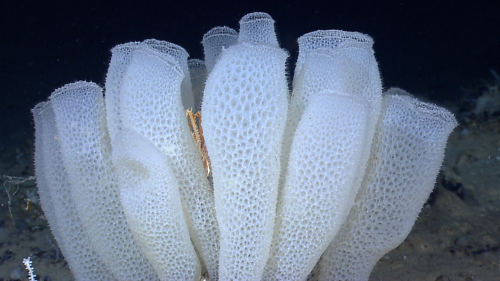
Leuconoids Leuconoid organization is the most complex of the sponge types and permits an increase in sponge size. In the leuconoid design, the surface area of the food-collecting regions with choanocytes is greatly increased; here the choanocytes line the walls of small chambers where they can effectively filter all the water present. The sponge body is composed of an enormous number of these tiny chambers. Clusters of flagellated chambers are filled from incurrent canals and discharge water into excurrent canals that eventually lead to an osculum.

oscula of the leuconoid canal system are seen at the edges of the plates. Unlike some other sponges, Mycale does not burrow into the coral skeleton and may actually protect coral from invasion by more destructive species. Pinkish radioles of a Christmas tree worm, Spirobranchus giganteus (phylum Annelida, class Polychaeta) also project from the coral colony. An unidentified reddish sponge can be seen to the right of the Christmas tree worm.
A sponge pumps a remarkable amount of water. Leuconia (Gr. leukos, white), for example, is a small leuconoid sponge about 10 cm tall and 1 cm in diameter. It is estimated that water enters through some 81,000 incurrent canals at a velocity of 0.1 cm/second in each canal. However, because water passes into flagellated chambers with a greater cross-sectional area than the entry canals, water flow through the chambers slows to 0.001 cm/second. Such a flow rate allows ample opportunity for food capture by choanocytes. Leuconia has more than 2 million flagellated chambers where food collection occurs.
After food is removed, the used water is pooled to form an exit stream. The exit stream, containing the entire volume of water that entered the sponge over the myriad incurrent canals, leaves the sponge through an exit pore whose cross-sectional area is many times less than the total cross-sectional area of all the incurrent canals. The relatively small size of the exit pore, together with the large volume of used water, produces a very high exit velocity. In Leuconia, all water is expelled through a single osculum at a velocity of 8.5 cm/second—a jet force capable of carrying used water and wastes far enough from the sponge to avoid refi ltering.
Some large sponges can filter 1500 liters of water a day, but unlike Leuconia, most leuconoids form large masses with numerous oscula, so that water exits from many local sites on the sponge. Most sponges are of the leuconoid type; leuconoid bodies account for most species within class Calcarea and are the most common types in other classes.
Useful External Links
- Phylum Porifera: Canal System in Sponges, Types of Canal Systems in Sponges, Functions of Water Current by STUDYANDSCORE
- Give a brief account on the canal system in sponges by Vedantu
- Canal Systems Encountered in Different Sponges by Biology Discussions